
Cancer is nowadays one of the leading causes of death worldwide. Early diagnosis is still one of the best strategies to improve the efficacy of clinical treatments. Recent developments at the frontier of research have focused their attention on the design of innovative contrast agents, endowed with high biocompatibility, sensitivity and specificity. The goal is to assess the presence of a pathology, especially cancer, with speed and precision, to provide targeted and timely treatments.
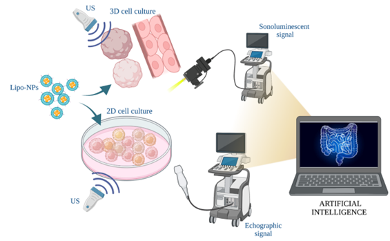
To improve the technologies for early tumor diagnosis, the TNHLab have patented an innovative system for the detection of optical signals and the formation of images deriving from cells and/or tissue (“METHOD AND APPARATUS FOR NANOPARTICLE- ASSISTED SONOLUMINESCENCE CONTRAST IMAGING IN MEDICAL DIAGNOSTICS, IN PARTICULARY ONCOLOGY” Priority N° 102021000005123 PCT IB2022/051802). The system includes an ultrasonic pressure waves generation unit, causing the cavitation of microbubbles and the consequent emission of sonoluminescent light increased by the presence of biomimetic nanoparticles (Lipo-NPs). These are characterized by specific optical properties capable of absorbing, scattering and modifying the light spectrum of sonoluminescence and give a specific signature to be detected by the system.
To optimize both the resolution and the detection depth of the diagnostic signals, in this project a system with a double imaging method is proposed: the sonoluminescence described above, generated by ultrasound radiation, and the echography, improved in precision, depth of penetration and specificity thanks to the presence of Lipo-NPs. The structure and chemistry of Lipo-NPs has been studied to incorporate the greatest number of air bubbles on their surface, thus increasing the cavitation nuclei responsible for sonoluminescence and increasing the echogenic contrast during ultrasound exposure; the nanometric size of the Lipo-NPs particles also allows their internalization in tumor cells, increasing the precision of the imaging system. Furthermore, the biochemistry used to prepare them increases their specificity and selectivity towards the cells/tissue of interest, in this case the colorectal or the breast cancer mass.
The reconstruction of the images deriving from the double imaging method makes use of the support of artificial intelligence, capable of recognising cells, their metastatic evolution and constructing a 3D image of the area. The resulting images will be of high resolution, precision and noise-filtered, having the possibility to be further implemented by clinical data in the future in order to consider the whole clinical setting of the patient.
The proposed technology can have a high impact on the health of cancer patients, increasing the capacity for early diagnosis of tumor masses, even for clinically complex cases to be addressed with current investigation techniques. This technology could open up revolutionary prospects at both technological and industrial levels, in the field of high-tech, high-resolution imaging techniques and digital tools in advanced diagnostics.

This project has received funding under PNRR – M4C2 – AVVISO 3277/2021 – NODES – ECS00000036 – PoC NanoZoom, title “Nanoparticles as innovative high-contrast agents resolution and depth for tumor imaging”